


Optical Digital Micrography, Scanning Microscopy, Confocal Micrography, and Flat-Bed Scanning |
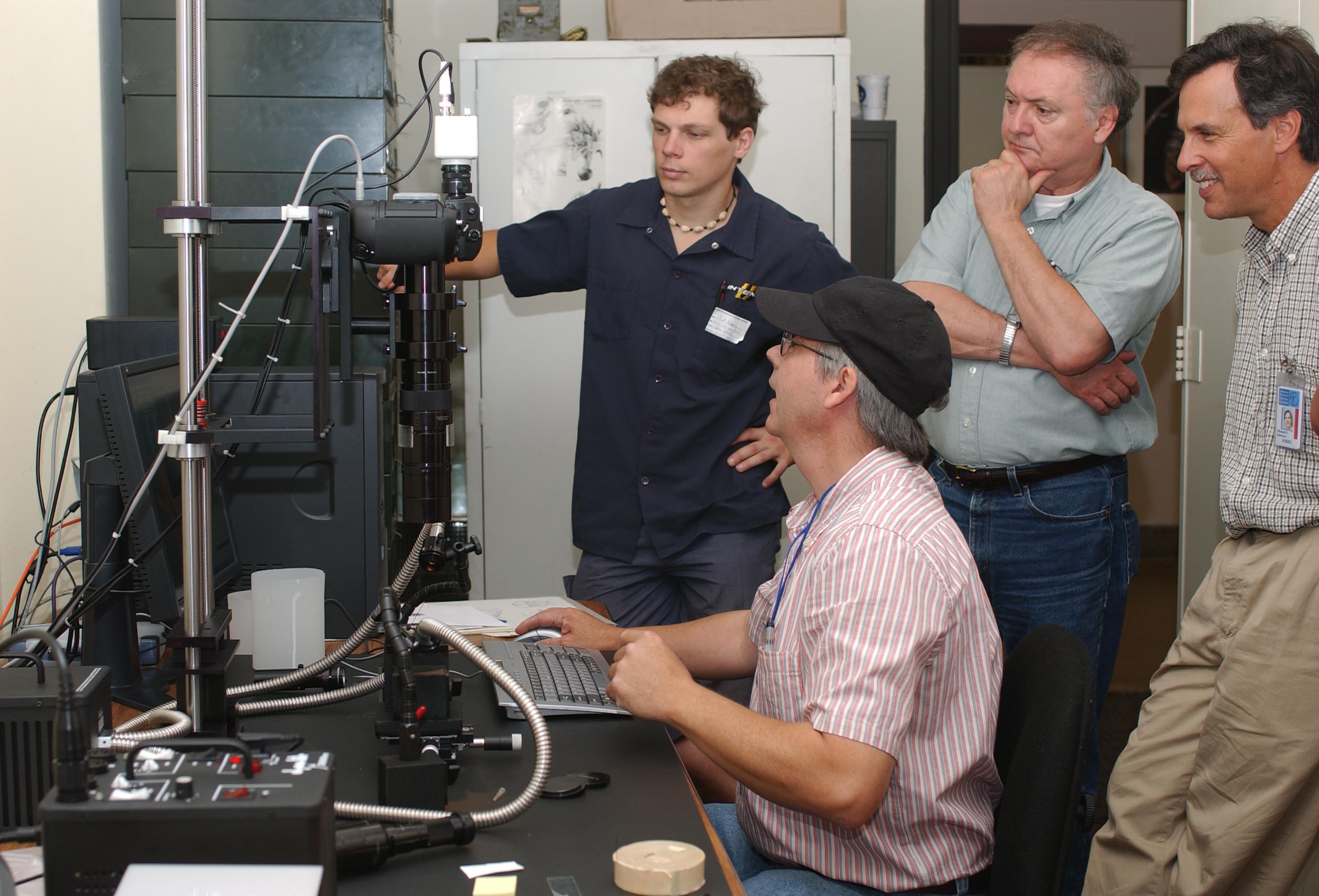 |
Optical Digital Micrography A core aim of the PBI project is to produce high quality digital images of as many taxa as possible, including holotypes. A review of available imaging system options led us to choose a Microptics-USA assembly, based on its unique combination of high depth of field optics, unique Microptics-USA stroboscopic lighting system, and real-time focusing, combined with other features such as a motor-driven lift. Equivalent units are stationed at the American Museum of Natural History, the Australian Museum, and the USDA Systematic Entomology Laboratory located in the National Museum of Natural History, Washington, DC. |
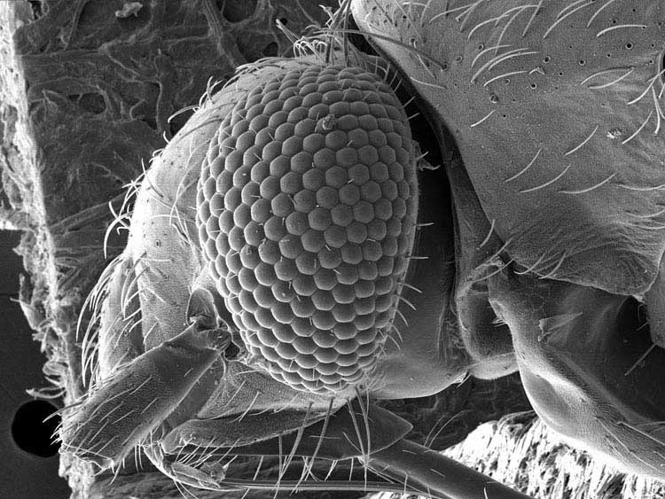 |
Scanning Microscopy The advent of digital scanning microscopy has moved this important arthropod imaging technique into the mainstream of systematic and morphological research. PBI participants at the American Museum of Natural History, the Australian Museum, Agriculture Canada, and the USDA Systematic Entomology Laboratory all have access to modern scanning electron microscopes that produce very high resolution digital images. This imaging technique allows for detailed structural investigation of setae, body surfaces, genitalic features, and external scent-gland features, among other characteristics. |
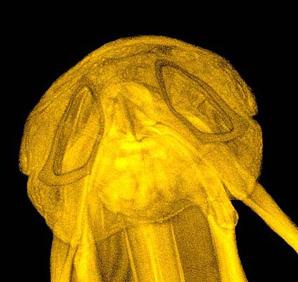 |
Confocal Microscopy Laser confocal microscopy offers an alternative technique for visualizing certain structures through the use of laser fluorescence imaging. PBI team members have applied this method as a way of improving our understanding of the relationships of complex genitalia structures, and intend to apply it to other structural systems. |
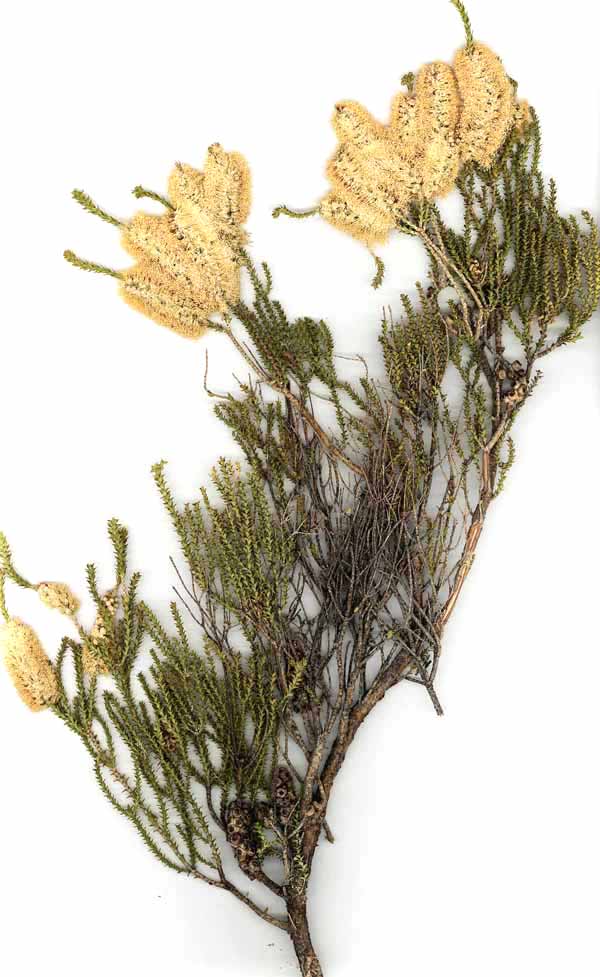 |
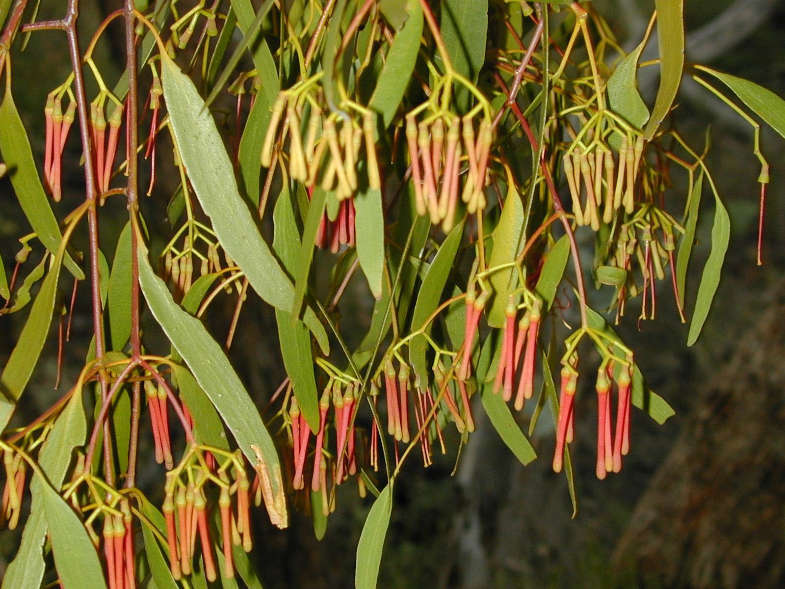
Flat-Bed Scanning of Host-Plant Vouchers Association of host plant voucher images with the specimen locality database provides an additional on-line source of information that precludes reference to the actual specimens when confronted with questions of accuracy of identification and plant condition (i.e., in flower, fruit, or sterile). We have applied the simple, but extremely effective, approach of using a high-resolution flat bed scanner to produce the plant-specimen images. With a limited amount of image manipulation, the shadows and other distracting elements can be removed from even the most highly three-dimensional specimens, such as those of Banksia spp. (Proteaceae) in flower or fruit. Such images, dating from fieldwork beginning in 1995 are being incorporated into the PBI website presentation. |
 |
Field Photographs Fieldwork associated with the PBI project dating from 1999 has relied heavily on the use of digital photography to document habitat types and the condition of hosts in life. These photos are displayed in conjunction with scanned host voucher specimens to provide an improved understanding of environmental conditions prevailing at the time of specimen capture. All such images will be made available through their association with the locality database. |