|
Standard Views and Imaging Protocols The imaging of terminals is done according to the protocols of the Manual for Standard Views
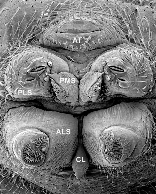
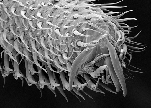
Morphology Each of the more than 500 spider species plus close
relatives (the
arachnid orders Amblypygi, Schizomida
and Uropygi)
included in the Tree of Life: Phylogeny of Spiders project will be documented
in hundreds of images. The primary tool for these morphological atlases will be
scanning electron microscopy (SEM). Using SEM, we can examine structures at a
much higher resolution than is possible with light microscopy. SEM is
especially suited to examining spinneret spigots and other fine details of
morphology. SEM imaging has played an increasingly critical part in the
discovery of new characters for phylogenetic analysis. Additional imaging will
be done using light microscopy with a series of images taken at different focal
planes combined using the software package Auto-Montage. Each taxon is assigned
to a clademaster, who is responsible for obtaining
specimens for both morphological and molecular analysis, and for completing the
series of images. Another important source of information is legacy data.
As of 2003, there were 68 cladistic analyses of spiders including morphological
data for three or more genera. Most of these were published or in press,
although some were in an advanced state of progress but not yet submitted
for publication. Taken together, these 68 matrices include members of
885 genera scored for over 4000 characters. Many of these characters
are similar to or duplicates of characters in other analyses, so a major
challenge was to synonymize characters across matrices. This matrix of legacy
data will be the starting point for the Tree of Life spider matrix.
Some legacy characters accepted as is for the Tree of Life project,
others will be modified to better suit the taxon sample, still
others will be excluded as not useful to address the deep scale questions
addressed by the Tree of Life project. New characters will also be added
based on the results of the SEM imaging and other observations.
|