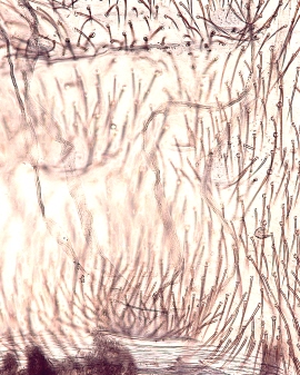
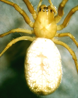
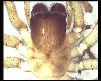
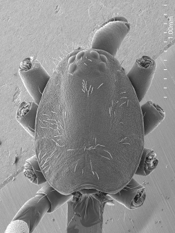
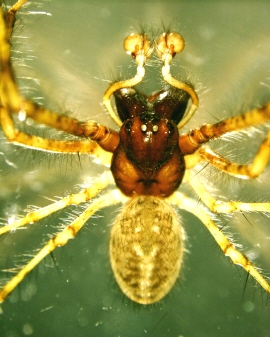
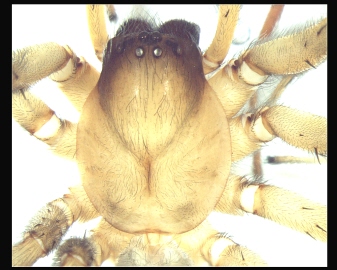
33. habitus (female): dorsal view (photo)
Tip: no legs
Manual for Standard Views
Please send corrections & nicer example images to [email protected]ID#. required
ID#. conditionally required
Preparation 1. Female in alcohol
|
33. habitus (female): dorsal view (photo)
Tip: no legs |
|
|
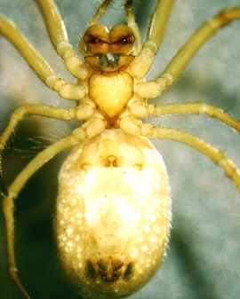
37. habitus (female): ventral view (photo)
Tip: no legs |
|
|
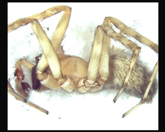
377. habitus with legs (female): lateral view (photo)
Tip: with legs |
|
|
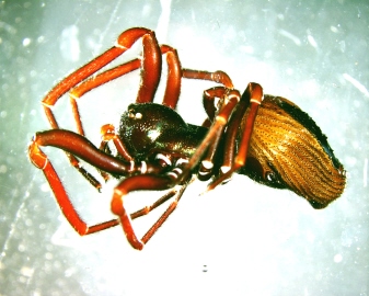
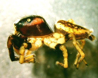
35. habitus (female): lateral view (photo)
Tip: no legs |
|
|
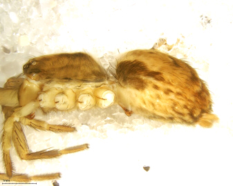
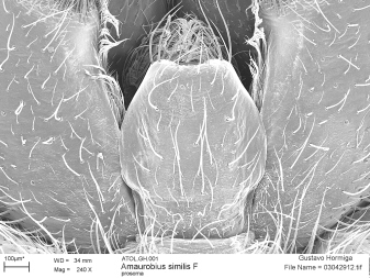
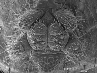
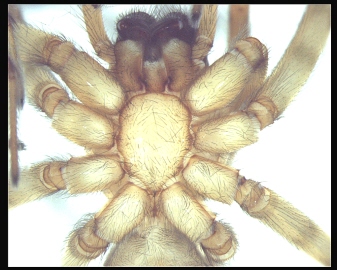
365. cephalothorax and base of appendages (female): ventral view (photo)
Tip: including coxae, trochanters, chelicera and pedicel |
|
|
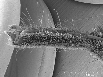
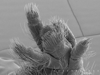
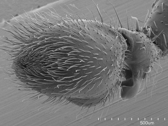
104. leg I (female): prolateral view (photo)
Tip: this can be imaged before mounting for SEM |
|
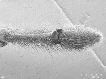
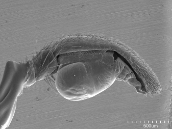
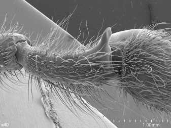
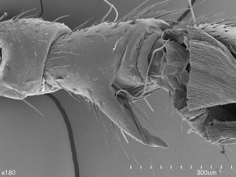
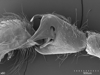
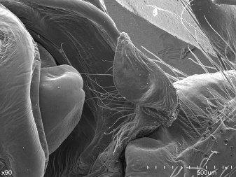
370. trochanter > leg I (female): ventral view (photo)
Tip: make sure the notch area is visible |
|
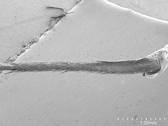
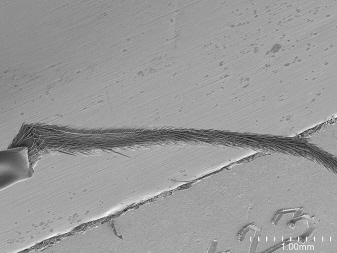
372. leg IV (female): retrolateral view (photo)
Tip: this can be imaged before mounting for SEM |
|
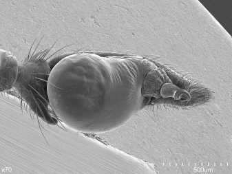
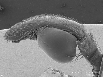
369. trochanter > leg IV (female): ventral view (photo)
Tip: make sure the notch area is visible |
Preparation 2. Carapace, endites, labium (female)
|
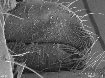
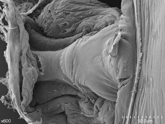
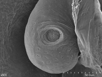
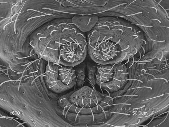
43. labrum (female): anterior-proper view (SEM)
Tip: elevated labra often require an oblique lateral view |
|
|
44. labium (female): ventral-posterior view (SEM)
Conditionally required: include if not adequately illustrated in SV 78 |
|
|
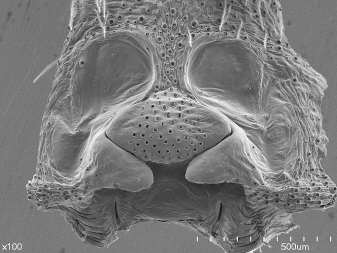
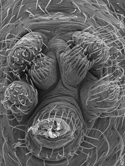
47. sternum (female): ventral view (SEM)
Tip: include pedicel
Zoom and check: if complex sclerites, then add SV 237 |
|
|
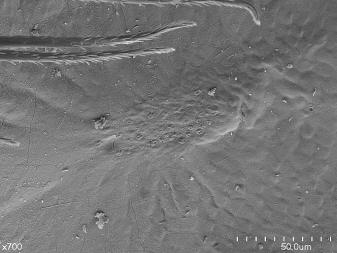
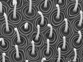
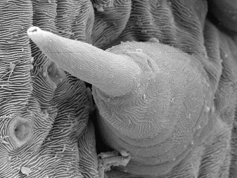
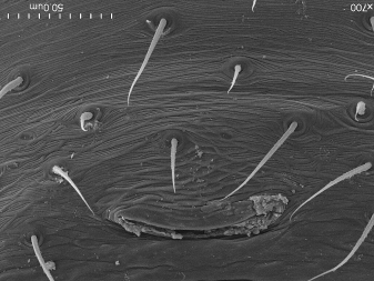
53. carapace (female): proper view, detail texture (SEM)
Conditionally required: include if not adequately illustrated in SV 67 |
|
|
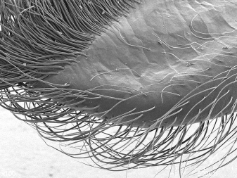
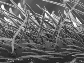
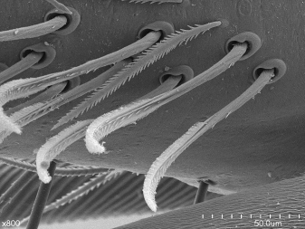
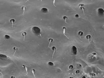
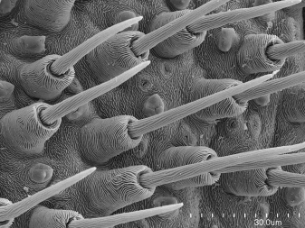
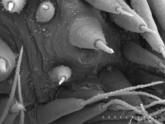
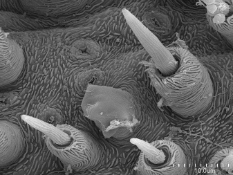
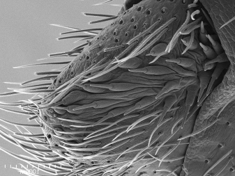
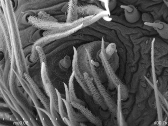
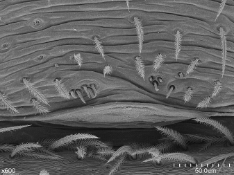
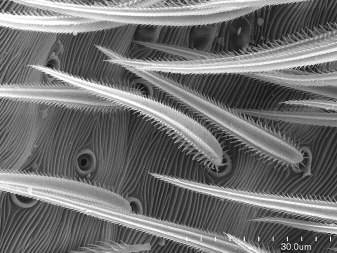
359. hairs carapace (female): proper view, closeup one seta (SEM)
Conditionally required: include if not adequately illustrated in SV 67 |
|
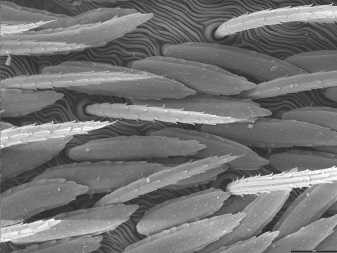
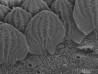
358. scales carapace (female): proper view, closeup one seta (SEM)
Conditionally required: include if not adequately illustrated in SV 67 |
May use standard legs and segments to illustrate setae, but include extra images if something different occurs elsewhere. |

|
|
|
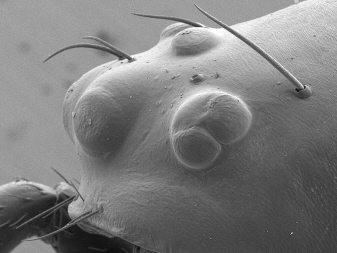
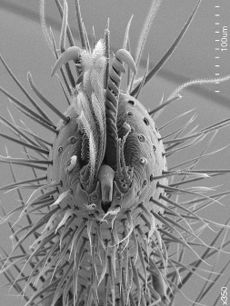
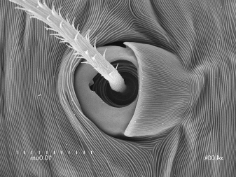
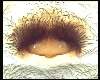
65. eyes (female): lateral view (SEM)
Tip: include all clypeus
Additional structures you may found there: if suprapalpal (?) glands present, add SV 6 |
|
|
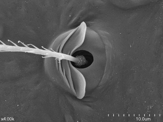
6. lateral pore pit (suprapalpal) > cephalic area (female): proper view (SEM)
When present! |
|
|
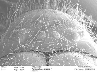
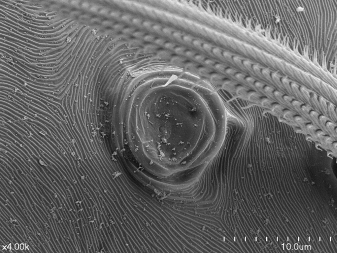
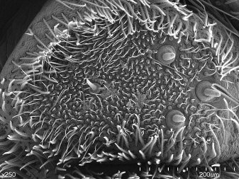
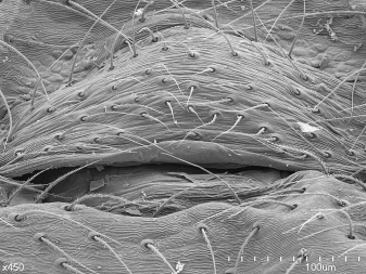
67. fovea > carapace (female): dorsal-proper view (SEM)
Tip: Fovea area, even if fovea not present
Zoom and check: ultrastructure of carapace cuticle (fingerprint, etc.), hairs, and scales; if not adequately resolved in this view, add SV 53, SV 358, SV 359 |
|
|
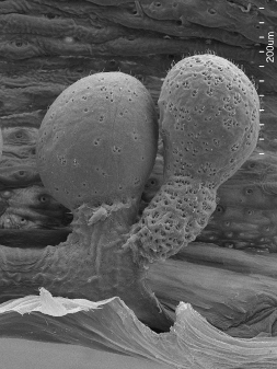
77. coxa-endite > palp (female): dorsal-anterior view (SEM)
Zoom and check: full extention of serrula, if teeth not well resolved add SV 79 |
The endites could go in the carapace preparation� 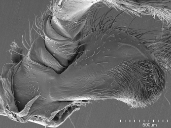
�or dissected with the palp |
|
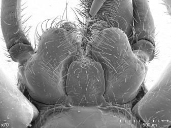
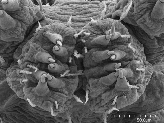
78. coxa-endite > palp (female): ventral-posterior view (SEM)
Tip: both left and right, including labium
Zoom and check: labium adequately resolved; if not, add SV 44 |
|
|
79. serrula > coxa-endite > palp (female): dorsal-lateral view (SEM)
Conditionally required: include if not adequately illustrated in SV 77
Zoom and check: serrula teeth well resolved |
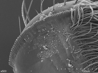
Document absence of serrula. |
|
234. pedicel (1st abdominal segment) (female): dorsal view (SEM)
Comments: Charles has asked for an oblique view; consider some examples and decide |
|
|
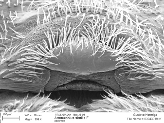
237. pedicel (1st abdominal segment) (female): ventral view (SEM)
Conditionally required: default view taken together with sternum in SV 47; add this view if complex |
Preparation 3. Chelicera (female)

When chelicerae are fused they could be mounted together |
|
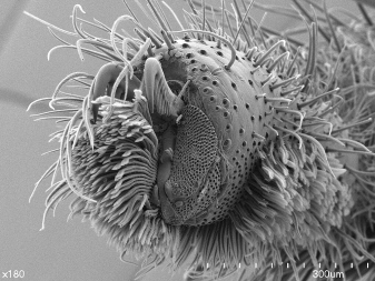
70. chelicera (female): posterior view (SEM)
Tip: If the venom gland comes out with the chelicera, mount and then take a whole image of chelicera plus venom gland |
Try to avoid closed fangs |
|
69. chelicera (female): ectal view (SEM)
Additional structures you may found there: stridulatory file, add SV 8 if necessary; cheliceral boss striae, if present add SV 7 |
|
|
7. cheliceral boss > paturon (female): lateral-anterior view (SEM)
When present! |
|
|
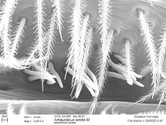
8. stridulatory files > paturon > chelicera > appendages cephalothorax (female): lateral-proper view (SEM)
When present! |
|
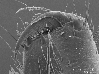
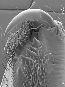
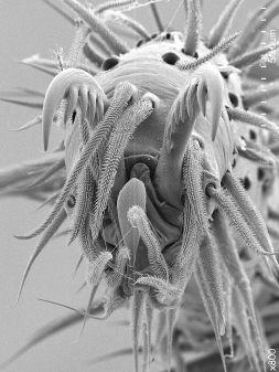
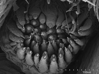
71. promargin > paturon (female): anterior-ventral view (SEM)
Comments: views differ in non-Araneomorphae |
Get all relevant setae in frame and in focus. 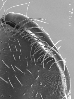
Include complete fang in frame. |
|
73. cheliceral gland (female): proper view (SEM)
Conditionally required: include if not adequately illustrated in SV 72 |
The cheliceral gland often needs a much higher contrast than the rest of the shots. |
Preparation 4. Palp (female)
|
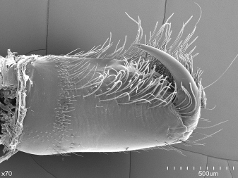
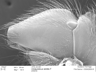
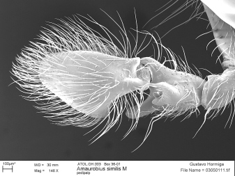
361. palp (female): prolateral view (SEM)
Tip: SEM of entire mount |
|
|

|
|
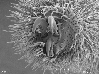
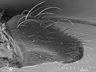
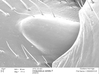
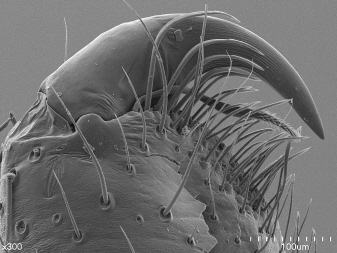
101. tarsus > palp (female): apical view (SEM)
Tip: retrolateral side shaved
Zoom and check: document absence of claw |
Document absence or reduction of claws. |
Preparation 5. Leg I, female
|
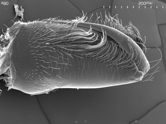
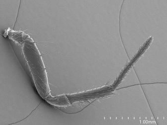
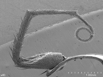
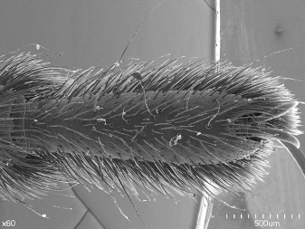
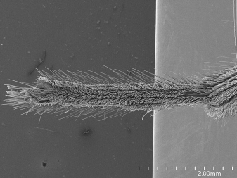
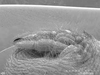
360. leg I (female): prolateral view (SEM)
Tip: general SEM of the entire preparation |
|
Include entire tarsus |
|
|
|
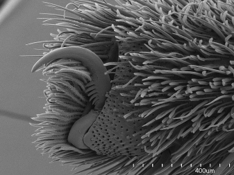
130. claws > leg I (female): apical view (SEM)
Tip: retrolateral side shaved, two images show teeth of all claws (or complete with extra images) |
|
|
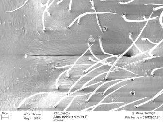
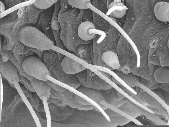
217. hairs appendages (female): proper view, closeup one seta (SEM)
Tip: generic; by default use tibia I lateral |
Look for diversity in setae. |
|
219. scales appendages (female): proper view, closeup one seta (SEM)
When present! |
|
|
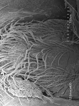
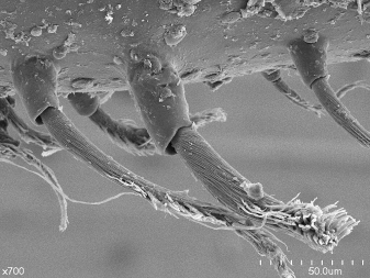
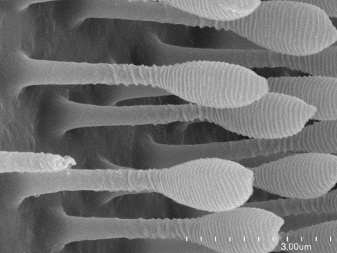
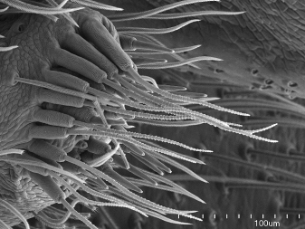
221. scopular setae (female): retrolateral-proper view, closeup one seta (SEM)
When present! |
Get well exposed base and tenent surface in focus. This image might be used as SV 219 as well, because the a scale is well figured along with the scopular seta. |
|
222. scopular setae tenent surface (female): proper view, closeup (SEM)
When present! |
|
|
15. claw tuft setae (female): retrolateral-proper view, closeup (SEM)
When present! |
|
|
16. claw tuft setae base (female): retrolateral-proper view, closeup (SEM)
When present! |

|
|
17. claw tuft setae tenent surface (female): proper view, closeup (SEM)
When present! |
|
|
223. chemosensory setae (female): proper view, closeup (SEM)
Tip: by default use cymbial tip or cymbial dorsal pad; check with some examples that e.g., tibial and cymbial setae look similar |
|
|
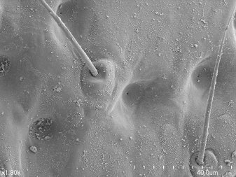
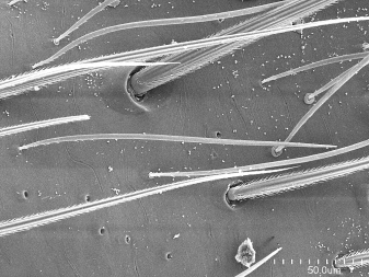
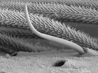
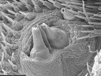
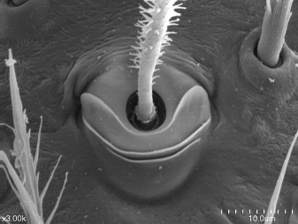
353. trichobothrial socket: proper view (SEM)
Tip: by default use female tibia I dorsal; make sure that the setal base is illustrated |
|
|
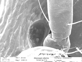
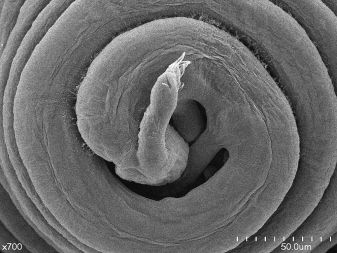
354. tarsal organ (female): dorsal-proper view (SEM)
Tip: generic; by default use tarsus I |
|
|
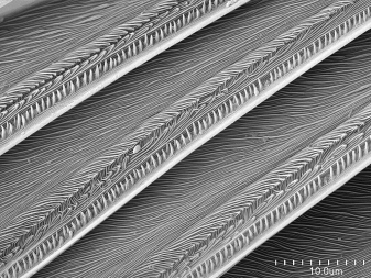
380. leg cuticle texture: proper view (SEM)
Conditionally required: include if not adequately illustrated in e.g., SV 353, SV 354
Zoom and check: Sometimes the cuticle texture varies in different parts of the leg (e.g., smooth on tarsus, but imbricate on tibia) |
The cuticle texture is often well illustrated together with trichobothria or tarsal organ
The cuticle texture is often well illustrated together with trichobothria or tarsal organ |
Preparation 6. Leg IV, female
|
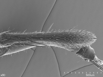
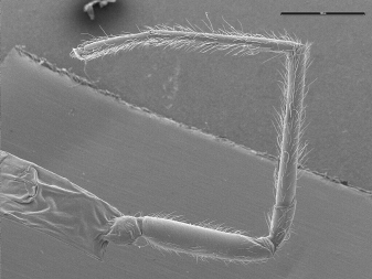
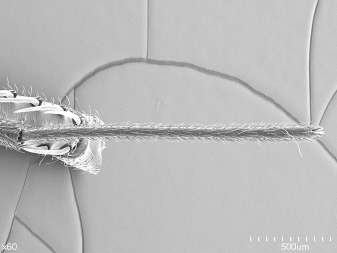
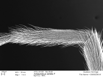
362. leg IV (female): retrolateral view (SEM)
Tip: general SEM of the entire preparation |
|
206. metatarsus > leg IV (female): dorsal view (SEM)
Zoom and check: metatarsal dorsal stopper may have oddities (Sparassidae) |
|
|
|
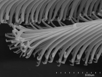
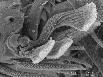
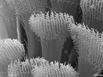
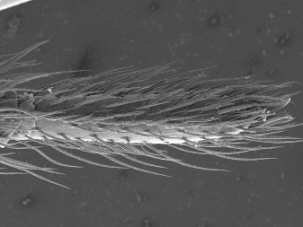
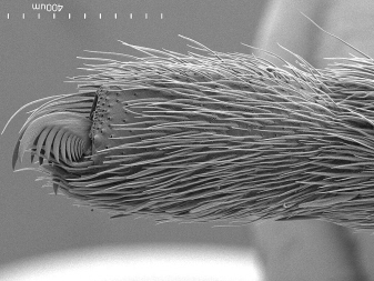
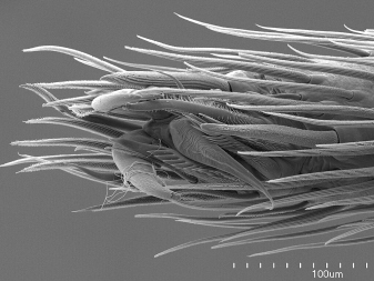
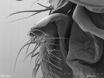
367. preening comb in metatarsus > leg IV (female): ventral view (SEM)
When present! |
|
|
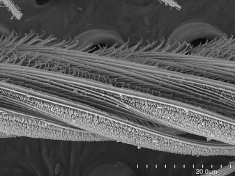
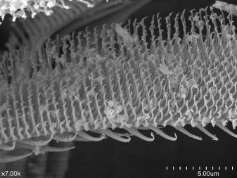
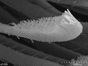
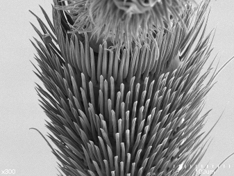
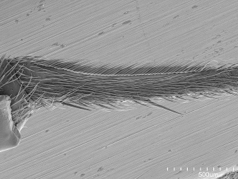
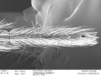
10. calamistrum (female): retrolateral-dorsal view, entire calamistrum (SEM)
When present! |
|
|
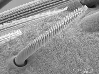
11. calamistrum (female): retrolateral-dorsal view, calamistrum sector several setae (SEM)
When present! |
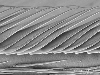
Check that tips and bases are in frame. |
|
|
213. tarsus > leg IV (female): ventral view (SEM)
Tip: include metatarsal tip
Zoom and check: preening comb resolved or add SV 367; tarsal comb resolved or add extra image (may need an additional, non-standard angle); sustentaculum resolved or add in SV 355, or directly as SV 13 |
Similar to this, but should include more of metatarsus tip. |
|
|
13. sustentaculum > tarsus > leg IV (female): proper view (SEM)
When present! |

|
|
14. tarsal combs > tarsus > leg IV (female): proper view, detail seta (SEM)
When present! |
|
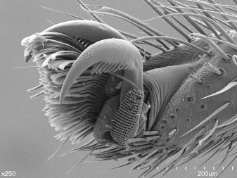
355. claws > leg IV (female): ventral view (SEM)
Tip: include modified setae close to claws
Zoom and check: accessory claws; if sustentaculum present and not resolved add SV 13 |
|
|
216. claws > leg IV (female): retrolateral-apical view (SEM)
Tip: retrolateral side shaved, two images show teeth of all claws (or complete with extra images)
Zoom and check: accessory claws |
|
|
214. claws > leg IV (female): apical view (SEM)
Tip: retrolateral side shaved, two images show teeth of all claws (or complete with extra images) |
|
Preparation 7. Epigyne and vulva trypsine digested
|
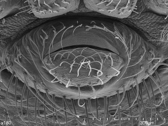
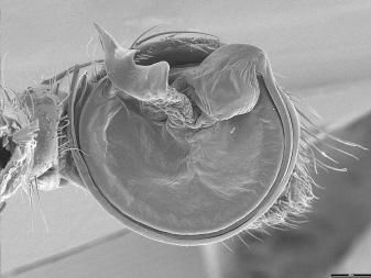
292. epigynum (female): ventral view (photo)
When present! |
|
|
293. epigynum (female): ventral view (SEM)
When present! |
|
|
290. epigynum (female): posterior view (SEM)
When present! |
|
|
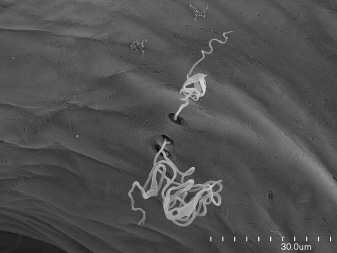
19. dictynoid pore > vulva (female): proper view (SEM)
When present! |
|
|
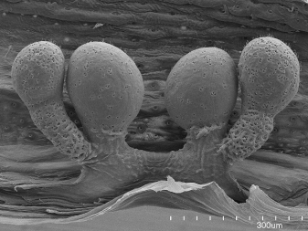
298. secondary spermatheca (SP2 or head) (female): proper view (SEM)
When present!
Zoom and check: including pores |
|
|
20. epigynal glands > vulva (female): dorsal-proper view (SEM)
When present! |
|
Preparation 8. Spinnerets female
One half is OK, but it is somewhat difficult to interpret spacing and general conformation. |
|
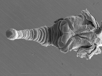
248. spinnerets (female): lateral view (SEM)
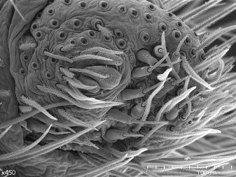
Tip: this is to illustrate the relative lengths of the spinnerets, especially the PLS |
|
|
251. AMS-cribellum-colulus (female): ventral view (SEM)
When present! |
|
|
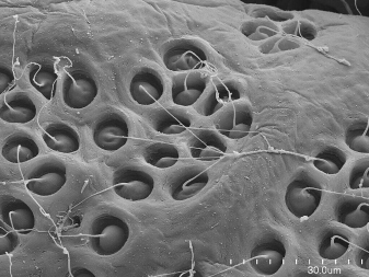
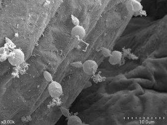
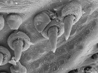
253. cribellar spigots (female): ventral-proper view (SEM)
When present!
Zoom and check: ultrastructural details of spigots |
|
|
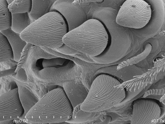
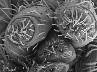
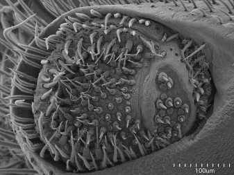
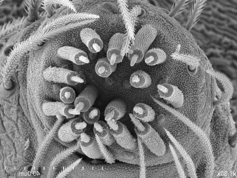
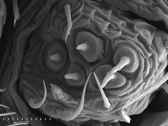
254. ALS spinning field (female): ventral-proper view (SEM)
When present!
Zoom and check: fine texture of spigots (shaft and base), tartipores, and spinning field cuticle, add extra images if necessary (SV 256, SV 258, SV 260) |
Shave distal article when necessary. |
|
256. maAmp > ALS spinning field (female): ventral-proper view (SEM)
Conditionally required: usually illustrated in SV 254 |
|
|
258. maAmp field > ALS spinning field (female): ventral-proper view (SEM)
Conditionally required: usually illustrated in SV 254 |

Get sensilla in focus. 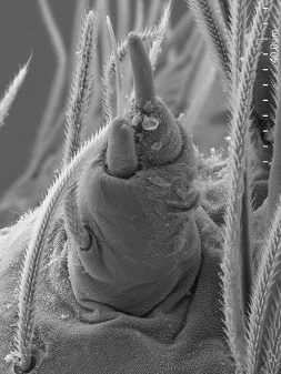
|
|
260. Pi > ALS spinning field (female): ventral-proper view (SEM)
Conditionally required: usually illustrated in SV 254 |
|
|
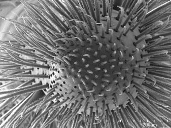
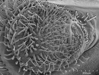
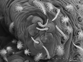
266. PMS spinning field (female): ventral-proper view (SEM)
Tip: entire spinning field or add extra images
Zoom and check: fine texture of spigots (shaft and base), tartipores, and spinning field cuticle, add extra images if necessary (SV 268, SV 270, SV 272, SV 273) |
|
|
268. miAmp > PMS spinning field (female): ventral-proper view (SEM)
When present! |
The same image will suffice to document both miAmp and Ac. |
|
270. Ac > PMS spinning field (female): ventral-proper view (SEM)
When present! |
The same image will suffice to document both miAmp and Ac. |
|
272. Cy > PMS spinning field (female): ventral-proper view (SEM)
When present! |
|
|
273. PC > PMS spinning field (female): ventral-proper view (SEM)
When present! |
|
|
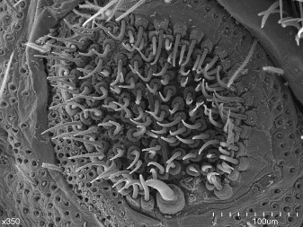
276. PLS spinning field (female): ventral-proper view (SEM)
Tip: entire spinning field or add extra images
Zoom and check: fine texture of spigots (shaft and base), tartipores, and spinning field cuticle, add extra images if necessary (SV 278, SV 280, SV 281, SV 283) |
Shave when necessary 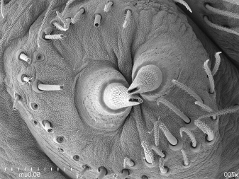
|
|
278. Ac > PLS spinning field (female): ventral-proper view (SEM)
When present! |
|
|
280. Cy > PLS spinning field (female): ventral-proper view (SEM)
When present! |
|
281. MS > PLS spinning field (female): ventral-proper view (SEM)
When present! |
|
283. PC > PLS spinning field (female): ventral-proper view (SEM)
When present! |
|
284. triad > PLS spinning field (female): ventral-proper view (SEM)
When present! |
|
|
288. anal tubercle (female): posterior-proper view (SEM)
Tip: one apical image should be sufficient, add more if needed |
|
Preparation 10. Male in alcohol
|
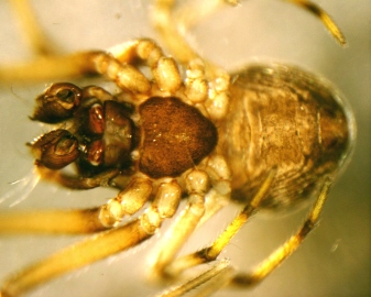
34. habitus (male): dorsal view (photo)
Tip: no legs |
|
|
38. habitus (male): ventral view (photo)
Tip: no legs |
|
|
36. habitus (male): lateral view (photo)
Tip: no legs |
|
|
378. habitus with legs (male): lateral view (photo)
Tip: with legs |
|
|
366. cephalothorax and base of appendages (male): ventral view (photo)
Tip: including coxae, trochanters, chelicera and pedicel |
|
|
|
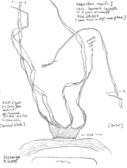
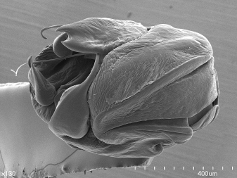
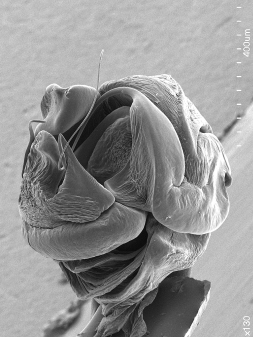
307. male palp and genitalia (male): ventral view, tarsus, bulb, tibia (photo)
Tip: this view can be replaced by a drawing (SV 306)
Zoom and check: degree of sclerotization of parts |

|
Preparation 11. Abdomen male
|
226. abdomen (male): anterior-dorsal view (SEM)
Zoom and check: illustrate sensilla, hairs and oddities around (especially on top of ) pedicel |
|
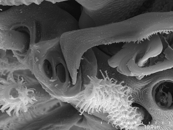
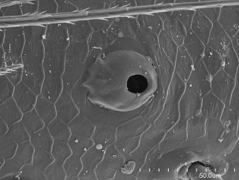
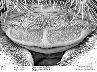
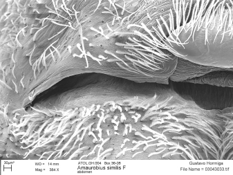
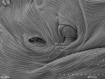
356. spiracle anterior booklung (male): ventral-proper view, left booklung (SEM)
Conditionally required: usually illustrated in SV 239 |
|
|
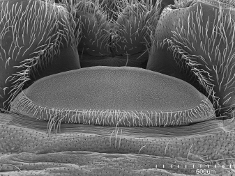
239. anterior booklung cover external surface (male): ventral-lateral view (SEM)
Tip: includes entire spiracle
Zoom and check: stridulatory surfaces; if spiracle not inlcuded, then add SV 356 |
|
|
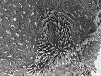
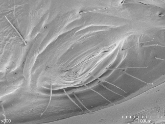
240. epiandrium (male): ventral-posterior view (SEM)
Zoom and check: if spigot origins not clear, add SV 18 |
Document absence. |
|
18. epiandric spigots (male): ventral-posterior view (SEM)
When present! |
|
|
373. spiracles > 3rd abdominal segment (posterior booklungs or tracheae) (male): ventral view (SEM)
Conditionally required: may be illustrated together with spinnerets in SV 249; shave area if necessary, use also to document absence of spiracle |
This view of the spinnerets may serve as well. |
|
|
255. ALS spinning field (male): ventral-proper view (SEM)
When present! |
|
|
267. PMS spinning field (male): ventral-proper view (SEM)
Tip: entire spinning field or add extra images |
|
|
277. PLS spinning field (male): ventral-proper view (SEM)
When present! |
|
|
285. triad > PLS spinning field (male): ventral-proper view (SEM)
When present! |
|
|
218. hairs abdomen (male): proper view, closeup one seta (SEM)
Tip: generic; default male, ventral; female ventral hairs from spinnerets mount is good as well |
|
|
220. scales abdomen (male): proper view, closeup one seta (SEM)
Tip: generic; default male, ventral; female ventral hairs from spinnerets mount is good as well |
|
Preparation 12. Palp, SEM (male)
|
352. male palp and genitalia (male): dorsal view, tarsus, bulb, tibia (SEM)
Tip: priority orthogonal to cymbium/tarsus longitudinal axis
Zoom and check: if not orthogonal to tibia longitudinal axis, or if tibia details not resolved, add SV 321; if trichobothria not resolved, add SV 28 |
Include full length of tibiae. |
|
349. male palp and genitalia (male): ventral view, tarsus, bulb, tibia (SEM)
Tip: priority orthogonal to cymbium/tarsus longitudinal axis
Zoom and check: if tibia details not resolved, add SV 324
Additional structures you may found there: modifications and special setae on cymbial tip |
|
|
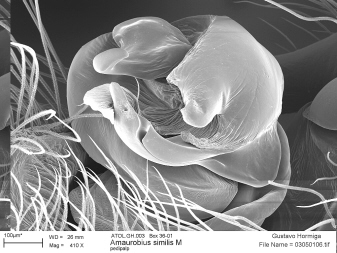
351. male palp and genitalia (male): prolateral view, tarsus, bulb, tibia (SEM)
Tip: can tilt slightly to get a better view |
|
|
350. male palp and genitalia (male): retrolateral view, tarsus, bulb, tibia (SEM)
Tip: can tilt slightly to get a better view
Zoom and check: if tibia details not resolved, add SV 323
Additional structures you may found there: modifications and special setae on cymbial retromargin, paracymbium (SV 29, SV 27) |
|
|
22. femoral thorns > femur > male palp and genitalia (male): prolateral view, closeup seta (SEM)
When present! |
|
321. tibia > male palp and genitalia (male): dorsal view (SEM)
Conditionally required: may be included in SV 352
Zoom and check: all processes
Additional structures you may found there: gland outlets, stridulatory areas |
|
|
324. tibia > male palp and genitalia (male): ventral view (SEM)
Conditionally required: usually included in SV 359 |
|
|
323. tibia > male palp and genitalia (male): retrolateral view (SEM)
Conditionally required: may be included in SV 350
Zoom and check: all processes
Additional structures you may found there: gland outlets, stridulatory areas |
|
|
28. trichobothria > tarsus-cymbium > male palp and genitalia (male): dorsal-proper view (SEM)
When present! |
|
|
326. tarsus-cymbium > male palp and genitalia (male): dorsal view (SEM)
Conditionally required: usually included in SV 352 |
|
|
29. retrolateral paracymbial processes (male): retrolateral-proper view (SEM)
When present! |
|
|
27. cymbial processes other than paracymbium > male palp and genitalia (male): proper view (SEM)
Conditionally required: usually included in general views |
For example, the thomisid "tutaculum". |
|
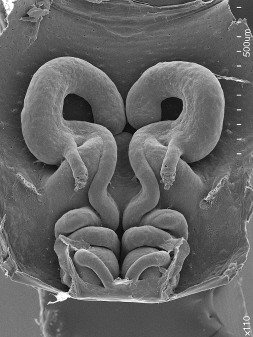
368. copulatory bulb (male): ventral view (SEM)
Zoom and check: all sclerites illustrated in this and other views of the bulb |
Sometimes the bulb is dissected and mounted apart. |
|
331. copulatory bulb (male): apical-proper view (SEM)
Tip: detail apical region, same preparation as SV 349 |
Sometimes the bulb is dissected and mounted apart. |
Preparation 13. Palp, expanded (male)
|
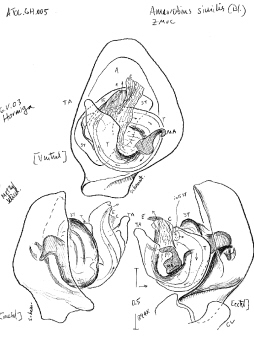
333. copulatory bulb (male): prolateral-proper view, expanded (photo)
Tip: only for expansible palps; can be replaced by an analytical drawing |
|
336. copulatory bulb (male): retrolateral-proper view, expanded (photo)
Tip: only for expansible palps; can be replaced by an analytical drawing |